Illuminating Endometriosis: Advanced Imaging Modalities and Their Clinical Impact
The Evolving Landscape of Endometriosis Diagnosis
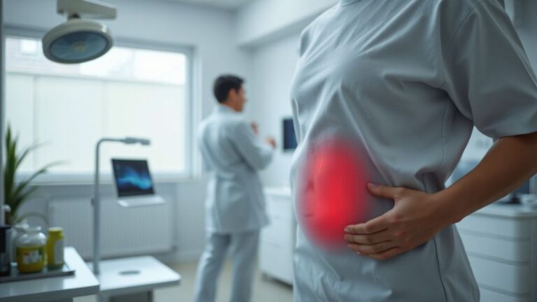
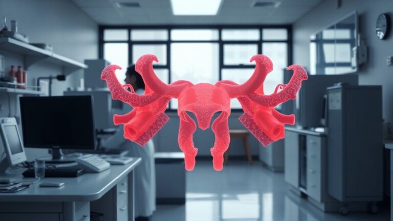
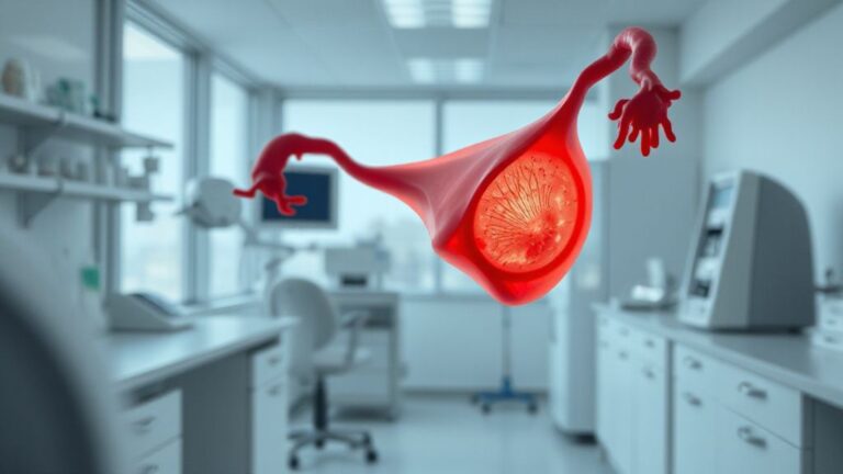
Endometriosis Imaging represents a critical frontier. This complex gynecological condition affects millions of women globally. It involves endometrial-like tissue growing outside the uterus. Consequently, it causes chronic pelvic pain and infertility. Traditional diagnosis relied heavily on laparoscopy. However, this is invasive. Therefore, non-invasive imaging techniques are paramount.
They offer earlier detection and better management strategies. Specifically, advanced modalities are transforming patient care. They provide detailed anatomical and pathological insights. This leads to more precise treatment planning. Beyond that, they reduce diagnostic delays. This is crucial for improving quality of life.
- ✓ Clinical Challenge: Distinguishing endometriosis from other pelvic pathologies.
- ✓ Diagnostic Goal: Achieving high sensitivity and specificity for various lesion types.
The quest for optimal diagnostic tools continues.
Current research focuses on integrating multiple imaging platforms. This aims to capture the full spectrum of the disease. It also addresses subtle presentations often missed. Consequently, patient outcomes are poised for significant improvement.
Ultrasound: The Foundation of Endometriosis Imaging
Transvaginal ultrasound (TVUS) remains a cornerstone of Endometriosis Imaging. It is widely accessible and cost-effective. Beyond that, experienced sonographers can identify typical endometriomas. These are ovarian cysts characteristic of the disease. Deep infiltrating endometriosis (DIE) is also detectable. This includes bowel and bladder involvement. Advanced ultrasound techniques enhance visualization.
antaranya, contrast-enhanced ultrasound (CEUS) can improve detection. It highlights vascularity of lesions. Doppler ultrasound assesses blood flow patterns. This helps differentiate endometriotic tissue from other masses. At the same time, 3D ultrasound offers superior spatial resolution. It aids in mapping lesion extent and relationships. Specifically, these advancements allow for more confident diagnosis pre-operatively.
This reduces the need for exploratory surgery in many cases. Consequently, patients benefit from timely interventions.
Magnetic Resonance Imaging (MRI): Unveiling Deeper Insights
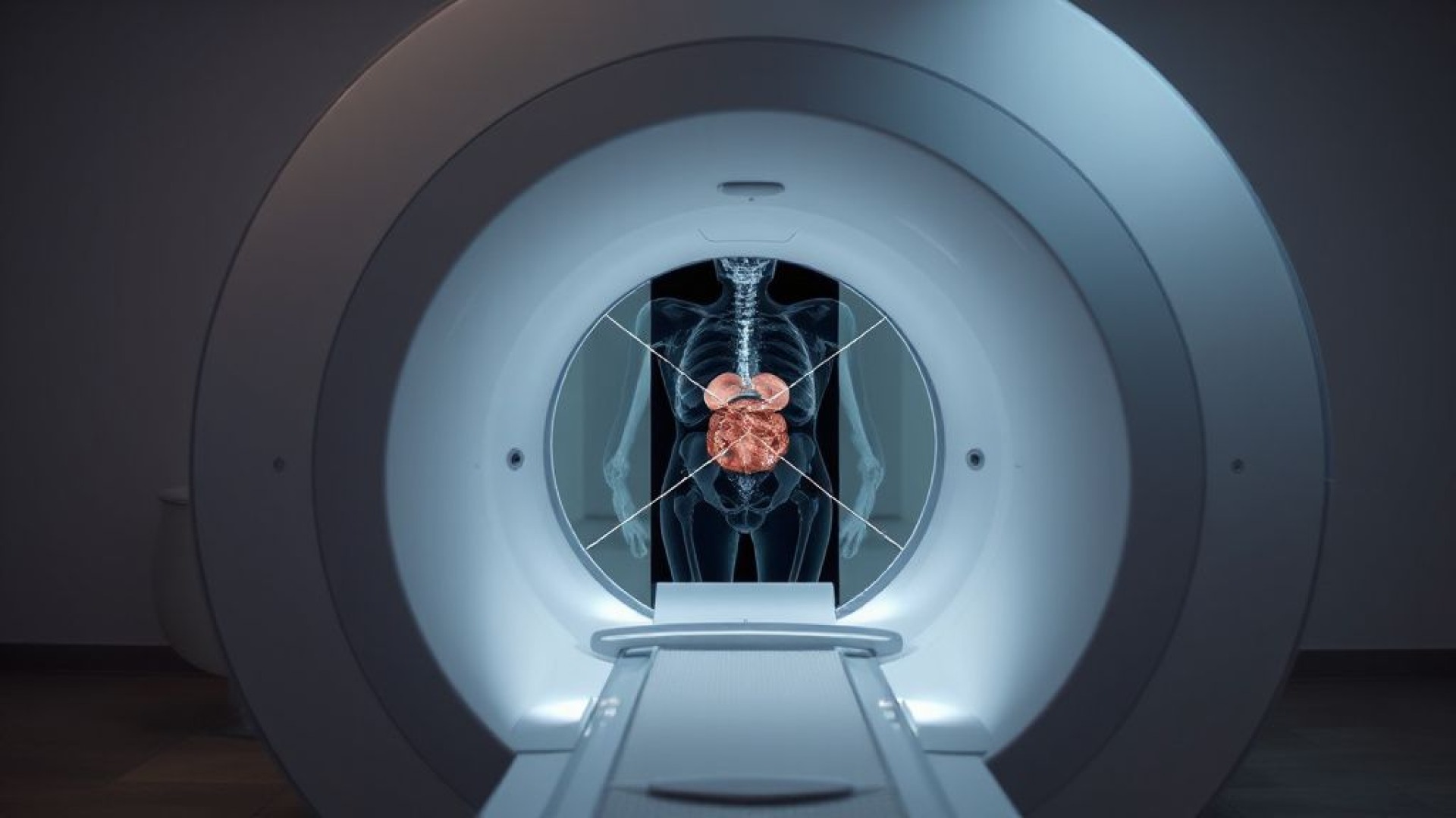
Magnetic Resonance Imaging (MRI) offers unparalleled soft-tissue contrast. It is highly valuable for Endometriosis Imaging, especially for DIE. MRI excels at visualizing pelvic sidewall involvement. It also precisely maps rectovaginal septum and uterosacral ligament lesions. Specific sequences like T2-weighted imaging are key. They clearly delineate the extent of endometrial implants.
Diffusion-weighted imaging (DWI) can further characterize lesions. It helps differentiate active disease from scar tissue. Beyond that, functional MRI techniques are emerging. These explore metabolic and molecular changes. The clinical utility of MRI is undeniable. It provides detailed anatomical mapping. This is crucial for surgical planning.
Surgeons use MRI to anticipate challenges. They can better plan resection strategies. Consequently, operative morbidity can be reduced. The integration of specialized MRI protocols is essential.
Computed Tomography (CT) and Other Modalities
While less sensitive for superficial endometriosis, Computed Tomography (CT) plays a role. It is particularly useful for assessing complications. For instance, it can detect bowel obstruction or ureteral obstruction. It also helps evaluate distant metastases, though rare. CT’s strength lies in its speed and availability.
However, its soft-tissue resolution is inferior to MRI. Therefore, it is rarely the primary diagnostic tool for endometriosis itself. Other emerging techniques are also under investigation. Positron Emission Tomography (PET) imaging shows promise. It can potentially identify metabolically active lesions. However, its role in routine clinical practice is still limited.
The future of Endometriosis Imaging may involve multimodal approaches. Combining strengths of different technologies offers comprehensive assessment. This provides a complete picture of disease burden. Consequently, treatment decisions become more informed.
Clinical Utility and Patient Management Integration
The clinical utility of advanced Endometriosis Imaging is profound. Accurate imaging guides therapeutic decisions. It helps differentiate between medical and surgical management. For DIE, pre-operative MRI is often standard. It informs surgical approach and potential complexity. For suspected ovarian endometriomas, TVUS is typically sufficient. However, MRI can clarify ambiguous cases.
Beyond that, imaging helps monitor treatment response. It can assess the effectiveness of hormonal therapies. It also tracks post-operative recurrence. The integration of imaging findings with clinical symptoms is vital. A multidisciplinary team approach is ideal. This includes gynecologists, radiologists, and surgeons. They collaborate to interpret complex cases.
Consequently, personalized treatment plans emerge. This ensures optimal patient outcomes. The goal is to alleviate pain and restore fertility.
| Imaging Modality | Primary Strength | Key Applications in Endometriosis | Limitations |
|---|---|---|---|
| Transvaginal Ultrasound (TVUS) | Accessibility, Cost-Effectiveness | Ovarian Endometriomas, DIE Assessment | Operator Dependent, Limited for Deep Lesions |
| Magnetic Resonance Imaging (MRI) | Superior Soft Tissue Contrast | Deep Infiltrating Endometriosis (DIE), Pelvic Sidewall Mapping | Cost, Availability, Longer Scan Time |
| Computed Tomography (CT) | Rapid Assessment, Complications | Bowel Obstruction, Ureteral Involvement | Low Soft Tissue Resolution, Radiation Exposure |
| Contrast-Enhanced Ultrasound (CEUS) | Enhanced Lesion Vascularity | Improved DIE Detection, Differentiation | Requires Contrast Agent, Operator Skill |
Future Directions and Emerging Technologies
The horizon for Endometriosis Imaging is bright. Research is advancing rapidly. Artificial intelligence (AI) is poised to revolutionize analysis. AI algorithms can assist radiologists. They can detect subtle lesions missed by the human eye. Beyond that, AI can quantify disease burden more accurately. This leads to more objective assessments.
Novel imaging agents are also under development. These aim to specifically target endometriotic tissue. They could enhance detection sensitivity further. Biomarker discovery integrated with imaging is another frontier. Combining imaging data with molecular markers may offer a complete diagnostic picture. Ultimately, the goal is non-invasive, highly accurate diagnosis.
This will empower patients and clinicians alike. It promises earlier intervention and improved management. Consequently, the burden of endometriosis can be significantly reduced.
- ✓ Research Focus: Developing AI-driven image analysis tools.
- ✓ Clinical Innovation: Exploring novel contrast agents for enhanced lesion visualization.
The continuous refinement of imaging techniques is essential.
It ensures that diagnosis and management keep pace with scientific understanding. This proactive approach benefits countless women.
Endometriosis: Advanced Diagnostic Imaging Modalities and Their Clinical Utility
Clinical Summary: Advanced imaging modalities like MRI and specialized ultrasound significantly enhance the accuracy and utility of endometriosis diagnosis, leading to improved patient management and outcomes.
- ✚ Improved visualization of deep infiltrating endometriosis (DIE)
- ✚ Enhanced differentiation of endometriotic lesions from other pelvic pathologies
- ✚ Precise anatomical mapping for surgical planning
- ✚ Facilitation of non-invasive diagnostic pathways
⚠️ Clinical Precaution: Consultation with a qualified radiologist and gynecologist is essential for appropriate imaging modality selection and interpretation.
👉 Experience the visual edition: Watch the Web Story ⚡. Access verified research updates directly on zeudytech.com. Cross-reference technical records via National Institutes of Health (PubMed Clinical Database).